Apoptosis:
Introduction:
Cell Proliferation and Apoptosis:
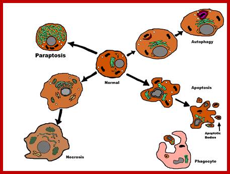
Repeated cell division and programmed cell differentiation is responsible for the development of a multicellular organism. Cell cycle events are themselves are programmed and genetically regulated. Pronounce Apoptosis as Apo-Ptosis not as Apop-tosis. In older organisms cells in tissues die for various reasons, one is injury that leads to death of cells by what is called necrosis. But certain injury at molecular level is deadly in the sense it can cause injury to other nearest neighboring cells, so cells have inbuilt sensing mechanism by which they initiate programmed cell death. If there is any loss of cells in a given tissue, for cell replacement, quiescent cells get activated to divide and redivide till the number of cells is averaged out. Exception to this is neuronal cells. Death of neuronal cells is permanent, and they are not replaced as in other tissues. But experiments in mammals and humans demonstrated later that new neurons are created in the central nervous system (CNS) in adults, although it seems to be restricted to some particular regions: granule cell layer of the olfactory bulb and dentate gyrus of the hippocampus. They come from neural stem cells (NSCs) that were preserved in the sub-ventricular zone during development. New cells in the nervous system wouldn't do any good. The whole nervous system is based on interneuronal connections, so adding an extra neuron would mess up these connections and alter both the functionality and the "stored" information. These new neurons seem to be local circuit neurons and interneurons (i.e. no long distance neurons). Death comes to cells in different forms so different names:
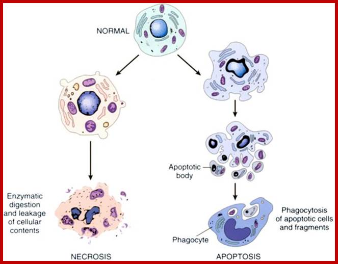
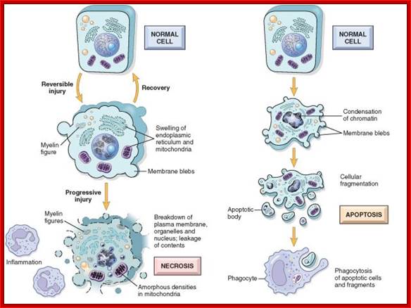
Necrosis: Cells get injured, cells get punctured where cells lyse extruding various injurious components, which cause severe damage to other neighboring cells, causing a widespread destruction; this is like carnage.
http://www.youthhunger.com
Paraptosis: Cells swell, develop large bubbles or vacuoles with liquid inside and die; this method of suicide is called parapoptosis. They don’t employ Caspases, which is hallmark of apoptosis. This method or most similar methods have been observed in yeast cells.
www.wikipedia.org
Autoschizis: It is a bizarre type of death. Cells develop crates inside and cell organelles escape from the cell and they are destroyed by some proteases that develop inside the cell. This happens when cancerous cells are treated with Vitamin-C and K.sub3. Normal cells remains unaffected, but many cells die because induced apoptosis, but substantial number of cancer cells die by autoschizis.
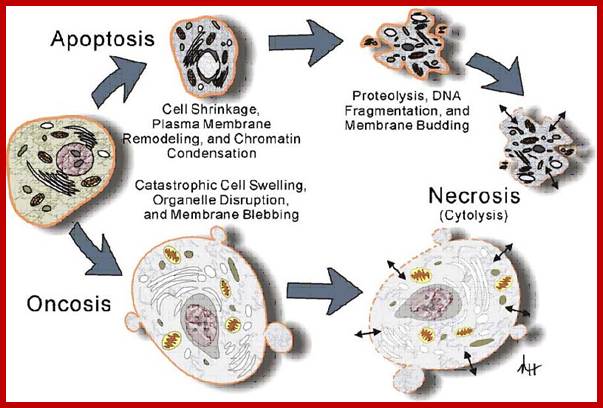
Oncosis: Cells expand by taking in lot of water in an uncontrolled manner. Soon proteins become denatured like cooked yolk proteins, then cells take excess calcium into cells and death follows.
Oncosis is the induction of necrosis by a nonphysiological event resulting in cell swelling as opposed apoptosis where there is cell shrinkage. Oncosis is induced by heat shock (56C or 42C), sodium azide (1% solution), Triton X-100 (0.01% solution) or drugs used to induce apoptosis at a higher concentration. Oncosis; In oncosis, annexin V is also externalized during the early phase of oncosis; the cells then rapidly proceed to cell death and losing membrane integrity, see figure. Oncosis is induced by heat shock (56C or 42C), sodium azide (1% solution), Triton X-100 or drugs used to induce apoptosis if used at higher concentrations can induce oncosis. http://www.icms.qmul.ac.uk
http://www.intechopen.com
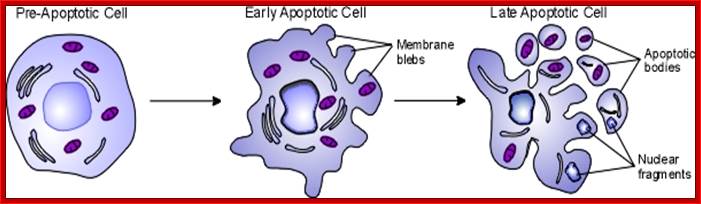
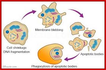
Apoptosis: Programmed cell death has certain morphological manifestations in contrast to cell death by necrosis and others mentioned above. On the contrary, Apoptosis or what is called -+programmed cell death, is stimulated by certain factors and leads to DNA fragmentation of 180 base pairs long (characteristic of apoptosis), intracellular organelles undergo fragmentation including nucleus and the cell collapses producing blebs and membranous vesicles. Most of these vesicles contain cellular components. When they are budded off they are recognized and engulfed by macrophages and consumed.

Fig-top-http://medicinembbs.blogspot.com; fig-bottom-http://www.utm.utoronto.ca

Apoptosis is essentially programmed cell death, or the common way that the body normally rids itself of cells that are not dividing properly. It is a normal feature of healthy organisms that is caused by a cell’s production of self-destructive enzymes, and occurs in the development of infants. However, apoptosis is shut off in cancerous cells – so damaged and/or mutated cells are not destroyed.
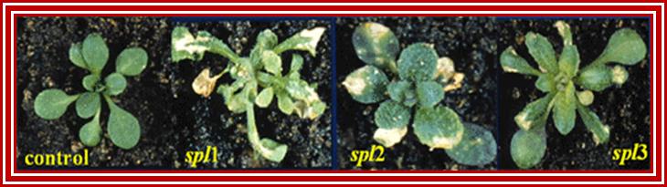
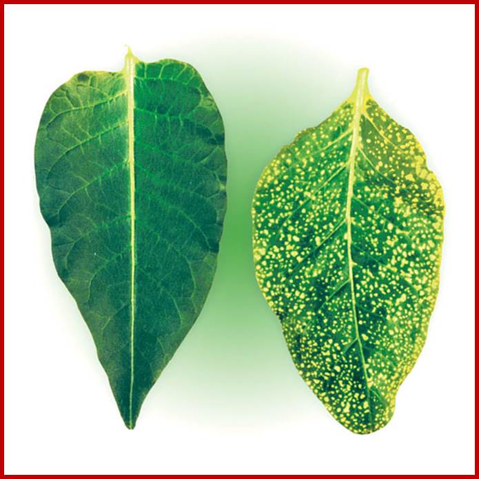
Figure shows plant cell death in leaves marked by their dried nature; https://www.quora.com; https://www.slideshare.net
Programmed cell death, mitochondria and plant hypersensitive response; http://www.nature.com/
The way it works is, cells release an intracellular apoptotic signal in response to a stress. There are two ways in which this signal is received: either by affecting mitochondrial function (intrinsic), or through directly passing the signal to the apoptotic mechanisms (extrinsic). The mitochondrion is the part of the cell that is responsible for aerobic respiration and for essentially making all the energy that your cells require to survive. If signals are received that alter its function, this tells the cell that it is essentially committing suicide and triggers the apoptotic mechanism. On the other hand, passing the signal directly onto the apoptotic mechanisms, in a process called signal transduction - which is basically like a relay race through the cell passing on a signal from one protein to another - by-passes the mitochondrial step. Either way, the end-result is the same, and each way can be reversed if cells no longer need to die. http://sphweb.bumc.bu.edu/ http://sphweb.bumc.bu.edu

https://cellbiology.med.unsw.edu.au
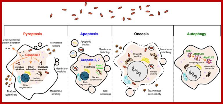
Pathogen-induced host cell death. Several forms of host cell death have been described during infection. The type of death the cell undergoes depends on the nature of the pathogen, pathogen load and site of infection. Pyroptotic, apoptotic, autophagic or oncotic cells display a distinct set of morphological and biochemical characteristics, some of which are shared. Whereas apoptosis and autophagy do not induce inflammation, the lysosomes of phagocytes that engulf apoptotic cells. During autophagy, pathogens are surrounded by autophagosomes and delivered to the lysosomes through autophagosome-lysosome fusion. Although apoptosis, pyroptosis and autophagy are generally beneficial cytokine release and escape of cytoplasmic content during pyroptosis or oncosis are highly inflammatory events. Pathogens are depicted as red ovals. During pyroptosis, pathogens (or pathogenic products) in the cytosol are detected by caspase-1-activating inflammasomes. During apoptosis, pathogens are contained within apoptotic bodies and digested in to the host, oncosis favors pathogen dissemination; Cell Death and Differentiation; K Labbé and M Saleh http://www.nature.com.
Development is genetically programmed and it goes through step by step and passes through various stages. During developmental stages, however, in a variety of animal and plant species, fully developed cells do undergo cell death.
· In plants, cell death is perfected in such a way, they leave structurally modified cells dead, but intact for specific functions, example most of the Xylem elements and sclerenchymal elements go through such process.
· In developing tadpole its tail is resorbed by apoptosis; the gradual degradation is not uninhibited invasive, once reaches a point, it stops.
· During human embryo development, the web like structure in between the fingers is gradually degraded by programmed death. Regression of human embryonic tail, nictitating membrane
· In the case of development of the nematode worm of Caenorhabditis elegans, cell divisions and differentiation leads to the formation of 1090 cells, of which 130 (1) cells die in a manner called programmed death of those cells only which are marked. This is an essential part of the development of this organism. It is not restricted to the worm; this phenomenon is spread over almost all multicellular organisms.
· Many neuronal and immune cells die a programmed cell death.
· In drosophila expression of rpr gene marks the cell for death. In moth during transition from pupa to insect, most of the muscle cells die. This death induced by the lack of ecdysone.
Cellular response to death signals leading to death:

Intrincic and extricic pathways; http://www.frontiersin.org
How do cells commit suicide; www.buzzle.com
Toxins induce cell death and apoptosis is induced to certain death signals;
http://www.unc.edu/;http://dc359.4shared.com/
· The cell death can be rescued by treatment with Actinomycin-D, (it is an Inhibitor of gene expression) which suggests that death requires certain genes to be expressed.
· In Munducta, Antherasa and Polyphemus (giant silk worm) during transition polyubiquitinin is expressed, and this targets protein for proteosome-mediated destruction.
· Cell death can also be due to cytotoxic T-cell, which attack marked cells and destroy by Granzymes. For example HIV infected cells do die by apoptotic process.
· The programmed cell death is predictable, defined in time and place. Death by this way is not passive but an active process involving gene activation.
· Dead cell can be detected by Acridine orange or Tryptan blue staining methods. Tryptan blue stains only dead cells. One can also use Propidium iodide, which is a DNA dye, which excludes dead cells. Cell death can also be monitored by the increased activity of polyubiquitination, increased activity of calcium dependent endonuclease activity, which cuts DNA into 180bplong fragments.
· Expression of receptors with death domain at cell surface is another determinant of death process..
Apoptic signals:
· The most common factor for cells to induce apoptosis is lack of essential growth factors.
· Ecdocyne deficiency in Moth.
· The Tumor Necrosis Factor (TNF) family comprises several ligands, such as the prototype TNF-, the Fas Ligand (Fas) and TNF-Related Apoptosis-Inducing Ligand (TRAIL/Apo2L).
· Thymocyte cells treated with glucocorticoids.
· Androgens in prostate cancer cells.
· Cells exposed gamma irradiation.
· Often cell are marked, example cytotoxic T-cells attack and kill target cells, which are immunologically marked.
· Irreparable damage to DNA. Internal factors such as Myc and
· p53 and its associated inhibitor proteins respond to such damages and initiated death..
· Any other stress factors can induce cell death; this amounts to molecular insult to the cell; “when you are abused to such an extent, what is the use of living, so commit suicide to avoid the
insult”.
· Some of the ligands like Fas-L and TNF a 1 are expressed as membrane bound proteins, but they are cleaved and released as soluble forms. Soluble forms of ligands are mostly produced by macrophages. Some of the Tumor Necrosis factors (TNFs) produced are pleotropic forms and they elicit many cellular responses through the binding to their respective cellular receptors.
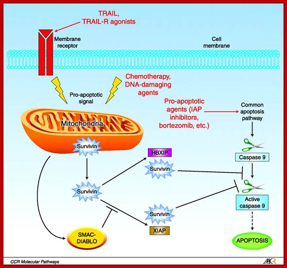
Function of Survivin as inhibitor of Apoptosis; Alain C. Mita,, et al, http://clincancerres.aacrjournals.org
|
Receptor |
Ligands |
Adaptors |
Targets |
|
|
Fas/CD95 Apo1 |
Fas-L Apo-IL |
FADD/ MORT1 |
Pro casp 8, |
Apoptosis |
|
TNF-R1 |
TNF-alfa |
TRADD, FADD |
Pro-casp 8 |
Apoptosis |
|
TN-R1 |
TNF-alfa |
TRDD/ RIP/TRAF2 |
MEKK, jun/Ap1 NFB |
Cell proliferation inflamation |
|
TNFR2 |
TNF alfa |
TRAF2/ TRF1 |
MEKK,IKK,NFkB |
Cell proliferation inflamation |
|
DR3,4,5 |
Trail APO-2L |
FADD |
Pro cas8 |
Apoptosis |
|
DCR1-3 |
TRAIL/APO2L |
None |
Decoy receptor |
Ligand sequestration |
TRDD= TNF receptor associate death domain.
RIP= Receptor interacting protein.
TRAF = TNF receptor associated factor.
TRAIL = TNF related apoptosis inducing ligands.
Which cells are marked for apoptosis?
1. Cells treated with carcinogenic chemicals, which express death signals.
2. Cells, which have no function. If there are excess cells than required they are subjected to death.
3. Cells that develop at improper places and at improper time they are induced for death.
4. Cells, which have completed their function and those having very harmful effect on other cells, are marked for death.
Effect of excessive apoptosis:
Many a times excess apoptosis takes place for wrong reasons, in such cases certain diseases develop, such as–insulitis, hepatitis, allergic encephalitis, Alzheimer disease, ischemic disease and many others.
Genes and the mechanism of apoptosis is discussed in the next chapter.